
128 Slice PET CT
Understanding PET scan
Positron Emission Tomography (PET) is a non-invasive molecular imaging technique that produces 3 dimensional images of functional/biochemical processes in the body.
In cancer and other diseases, biochemical or functional changes often precede structural changes. Hence PET identifies disease earlier than tests that rely on detecting a mass.
Biochemical/functional images of PET are combined with CT information, which can be obtained at the same sitting using our state of the art machine.
PET identifies abnormal biochemistry or disease. CT is used for precise anatomical localization and measuring (quantification) of the biochemical process.
Applications of PET
- Detection of cancer.
- Determination of whether the cancer has spread.
- Assessment of the effectiveness of a treatment plan, such as chemotherapy.
- Determination of whether a cancer has returned after treatment.
- Identification of areas of the heart muscle that would benefit from a procedure such as angioplasty or bypass surgery (in combination with a myocardial perfusion scan).
- Evaluation brain abnormalities, such as tumours, memory disorders and seizures and other central nervous system disorders.
How does a PET CT work in Oncology
Cancer cells grow at a fast rate utilizing glucose. Faster their growth, usually, higher is the rate of glucose consumption.
A radioactive form of glucose (FDG) is injected intravenously and the regional glucose metabolism of the whole body is mapped.
The nuclear medicine physician interprets the scan with the help of the CT scan for precise localization of cancer.
There are non-oncological applications as well of FDG PET scan in cardiac, neurological and infective conditions.
Preparing for the scan
- You should be fasted for at least 6 hours. Only water is allowed. No chewing gum, guthka, pan etc.
- All medications may be taken as usual. If they cannot be taken on empty stomach please contact your doctor.
- If you are a diabetic it is important that you consult your doctor in advance. Your blood sugar should be less than 150 mg/dL before the PET scan. If you are in any doubt contact the nuclear medicine department well in advance before booking an appointment.
- If you think you are or could be pregnant, please consult your doctor. PET scans are preferably avoided if you are pregnant.
- If you’ve ever had an allergic reaction to iodine, or if you have asthma, tell the doctor/ nurse. The doctor may prescribe special medicine for you to take before the test.
- If you are a large person or have a fear of enclosed spaces (claustrophobia), tell your doctor before the day of your PET/CT exam. The doctor can prescribe medicine to help you relax.
What will you bring with you and what you will be wearing?
- Please bring the plates/films of all relevant investigations like X-ray, CT, MRI or PET scans along with photocopies of the typed reports.
- Please bring the details of previous treatment related to the condition you are being scanned for (surgery, chemotherapy or radiotherapy). It is important that we have the right dates.
- Remove all jewellery or metallic objects at home before coming for your scan.
- Please maintain punctuality as FDG decays with time. If you need to cancel your appointment, do so at least 24 hours prior to your scheduled time.
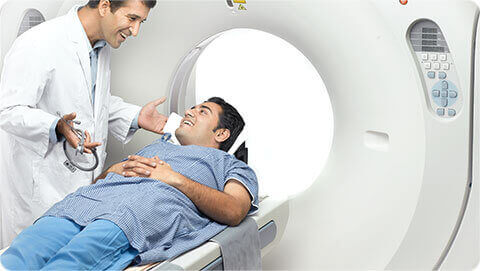
How will the scan be done?
- Every PET exam is different, but please expect to be in the dept for at least 2-1/2 hours
- On arrival, after registration, the nuclear medicine physician will explain the procedure and an IV canula
will be inserted by a technologist/caregiver. - A small amount of FDG will be injected through the IV access (painless).
- You will need to take oral contrast.
- After waiting for around 45-60 minutes, during which you must not engage in physical activity or talking, images will be acquired.
- It takes around 30 minutes for the scan to be performed. However if you need a contrast enhanced CT scan, additional time is
required for that.
What happens after the scan
- The physician in charge will review your PET scan and if required, will perform an additional contrast enhanced CT scan.
- You may eat, drink and do things (including driving) normally after the scan.
Is Radiation harmful?
Philips Time-of-Flight (TOF) PET system reduce the radiation exposure by more than 50% as compared to conventional PET/CT system. The overall exposure is small and acceptable.
The machine performing PET
We at Apollo Multispecialty Hospitals are committed to employing the latest technology for the benefit of the patients.We are equipped with one of the most technically advanced scanners, Philips Ingenuity-128 PET/CT system. It is the first system in the India to have 128 slice Ingenuity CT with Astonish 3rd generation time-of-flight (TOF) technology with the following unique advantage:
- A twofold higher PET sensitivity due to 3rd Generation TOF technology.
- An improved image quality by 30% noise reduction.
- Exceptional image quality at 50% less radiation (allowing half dose imaging).
- A unique open gantry design, resulting in more patient comfort.
- A 50% reduction of radiation and 30% improvement of image quality by Ingenuity CT.
- A 15% reduction in contrast toxicity.
The importance of PET imaging has increased immensely in recent years for diagnosis, staging and restaging for various cancer types.
- Non-Hodgkin lymphoma
- Hodgkin’s disease
- Gastro-intestinal stromal tumours
- Colorectal cancer
- Melanoma
- Breast cancer
- Head and neck tumours
- Evaluation of pulmonary nodules
- Gynaecological malignancies
- Thyroid carcinoma resistant to iodine therapy
- Metastatic disease with unknown primary cancer